The German physicist Alexander Behm (1880-1952), who taught at the Technical academy of Vienna from 1905 to 1913, discovered, in 1913, the principle of electroacoustic measurement of water. This depth-sounder technology (Echolot), still in use today, forms the basis of nautical navigation.
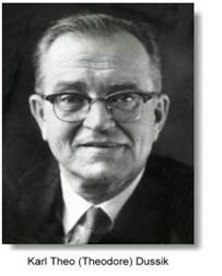
The Austrian neurologist Karl Th. Dussik (1908–1968) was the first doctor to use ultrasound for diagnostic purposes. Together with his brother Fritz, a highfrequency physicist, they tried to measure ultrasound beam absorption through the skull, hoping to obtain a picture of the brain. In 1942, he published the so-called “hyperphonography“ method.
„Über die Möglichkeit, hochfrequente mechanische
Schwingungen als diagnostisches Hilfsmittel zu verwerten“ >>, Karl Th. Dussik in: Zeitschrift Zeitschrift für die gesamte Neurologie und Psychiatrie 174 (1942):153-168.
Karl C. Ossoinig, who subsequently worked in the USA, at the University of Iowa City, was among the first to use ultrasound for ocular diseases. In Vienna during the early 1960s, i.e. the era of ultrasound’s discovery, he adapted the technology for diagnostic use in the eye. In this era, one had to use a water-bag to diagnose an eye tumour. Thus, the patient had to put on a fluid-filled diving mask. „Zur Ultraschalldiagnostik der Tumoren des Auges“ >>, Karl Ossoinig in: Klinische Monatsblätter für Augenheilkunde 146 (1965):321-37.
Alfred Kratochwil (*1928), pioneer of gynecologic ultrasound, by chance, was in the audience when a neurosuregon gave a lecture about using ultrasound to diagnose intracranial bleeds. Inspired by this, Kratochwil embarked on a longterm study and started a co-operation with the Kretztechnik Company. Using ultrasound equipment that was developed to check the railroad track, he was able to detect the placenta. Kratochwil introduced not only interventional ultrasound, but is also considered the father of modern sterile technique, both in diagnostic and therapeutic settings.
„Der früheste Nachweis der fetalen Herzaktion durch Ultraschall“ >>, Alfred Kratochwil und L. Eisenhut in: Geburtshilfe und Frauenheilkunde 27 (1967): 176-180.
Kretztechnik Company – from A-Mode Technique to 3D-Ultrasound: In 1947, Engineer Paul Kretz, from Upper Austria, started a small business. From 1953, the company developed ultrasound equipment used to check various metals, for eg. iron railroad tracks, brewery cauldrons. Soon, they became specialist in the development and production of ultrasound equipment intended for medical use. In 1989, the company presented the first 3D-ultrasound unit, worldwide, at a radiology conference held in Paris.
Colour-coded Doppler Sonography made it possible to distinuish veins from arteries, based upon the pattern of bloodflow; and to detect heart valve dysfunction and heart muscle defects. In order to process the data generated by the new generation of ultrasound units, computer development took off during the 1980s. This movement was led by B. Schwaighofer, P. Hübsch und F. Kainberger, all of whom were part of the Department of Diagnostic radiology, affiliated with the 2nd Medical University of Vienna. „Farbkodierte Doppler-Sonographie bei Nierentransplantaten“ >>, B. Schwaighofer, Hübsch P., Kovarik J., Frühwald F., Kainberger F., Barton P. in: Röfo - Fortschritte auf dem Gebiet der Röntgenstrahlen und der bildgebenden Verfahren 149 (1988):193‐196.
Wolfgang Buchberger, from the Radiology Institute at Innsbruck University, was the first to use ultrasound to examine the peripheral nerves (Neurosonography). It is due to his investigation of nerves of the carpal tunnel, that we can evaluate the status of various nerves today. „Carpal tunnel syndrome: diagnosis with high-resolution sonography“ >>, Wolfgang Buchberger, Judmaier W., Birbamer G. et al. in: American Journal of Roentgenology 159 (1992):793–798.